
ARVC WEB PAGES ARVC/ARVD
Pathology:St.Georges Hospital Medical School, London U.K.
Dr.Mark Norman
As can be seen here fat shown as yellow is replacing the normal heart muscle (brown). The fat tends to spread inwards from the outer surface of the heart ( the epicardium). When we look under the microscope we see fat and scar tissue the pathological hallmark of ARVC fat and fibrosis replacing the right ventricle. We also now recognise within ARVC families fat replacing the left ventricle also. It is presumed that the same genetic causes both variants of the disease.
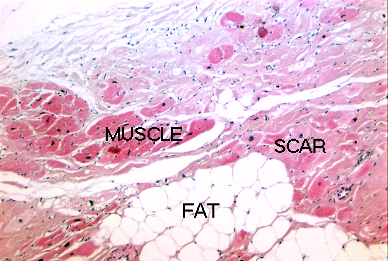 Post mortem or biospy specimens of tissue have a characteristic finding for ARVC. There is both FAT and FIBROSIS (scar tissue) replacing the muscle cells (See picture below). In normal hearts a quantity of fat replacing the heart muscle is seen. For ARVC to be diagnosed with certainty there must also be scar tissue. Muscle is seen as bright pink, scar tissue paler pink, and fat as clear spaces between the muscle cells and scar tissue.
![]()
Links to: